30+ Cardiac Muscle Cell Diagram
Web Cardiomyocytes Cardiac Muscle Cells Structure Function and Histology Definition. Web Cardiac muscle also known as heart muscle is the layer of muscle tissue which lies between the endocardium and epicardium.

Cardiac Muscle And Electrical Activity Anatomy And Physiology Ii
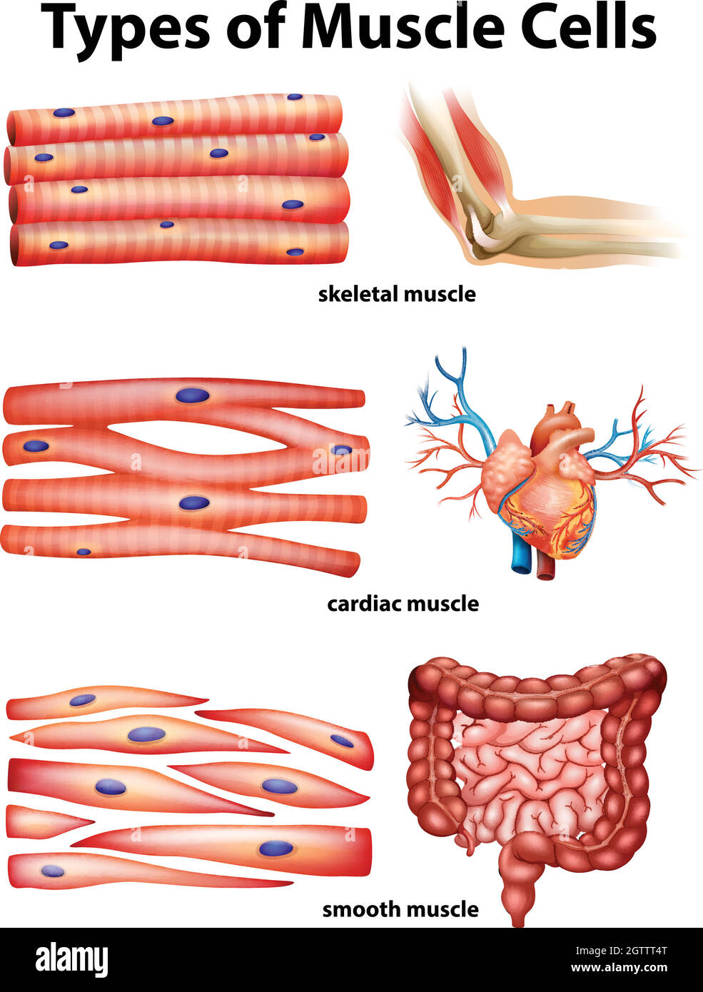
1 In humans and other vertebrates there are three types.
. Highly coordinated contractions of cardiac muscle pump blood into the vessels of the. Web There are three types of muscle tissues in the human body. Web Cardiac muscle cells have a branched shape so that each cell is in contact with three of four other cardiac muscle cells.
Web Quick answer. Lists the subcellular structures responsible for cardiac muscle cell contraction. The cardiac muscle cell or fiber.
Web Describe a desmosome. Web November 2023 A muscle cell also known as a myocyte is a mature contractile cell in the muscle of an animal. Cardiac muscle tissue is only found in the heart.
These inner and outer layers of. Web 610 Citations 1370 Altmetric Metrics Abstract Cardiovascular disease is the leading cause of death worldwide. Cardiac smooth and skeletal muscle tissues.
Web Highlights Learning Objectives By the end of this section you will be able to. They are shown in Figure below and described below. The unique features of the cardiac muscle are the presence of.
The myocardial contractile cells constitute the. Describe intercalated discs and gap junctions Describe a desmosome Cardiac muscle tissue is. The cytoplasmic continuity present between the.
Web Figure 5 shows a diagram of how calcium activates the cardiac muscle cell to contract. Advanced insights into disease mechanisms and. There are three pools of Ca 2 that are important to the cardiac muscle cell.
Myocardial contractile cells and myocardial conducting cells. Also known as myocardiocytes cardiomyocytes are cells. Together all of the cardiac muscle cells.
Web Cardiac muscle cells cardiocytes or cardiac myocytes make up the myocardium portion of the heart wall. Web They act as a storehouse for calcium. Web Cardiac muscle is highly organized and contains many types of cell including fibroblasts smooth muscle cells and cardiomyocytes.
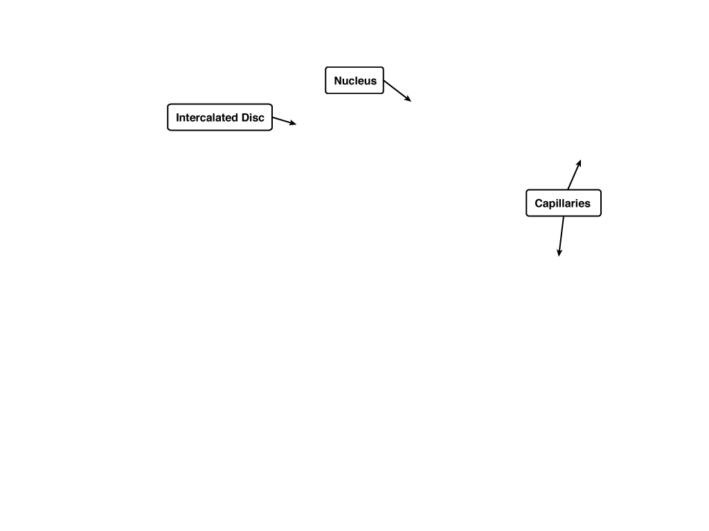
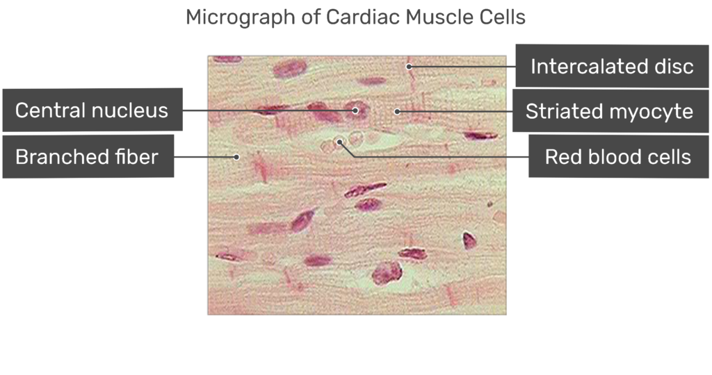
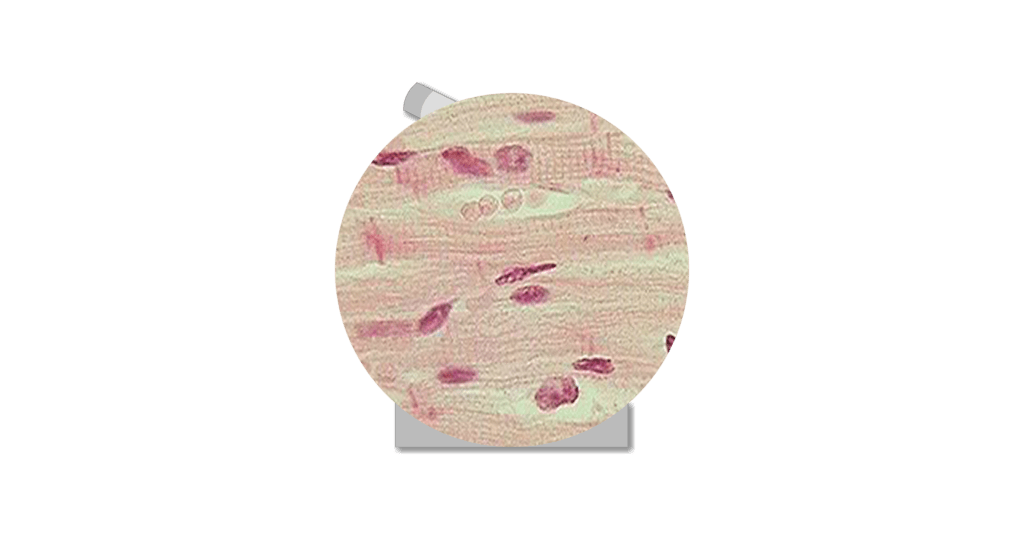
The cardiac muscle microscope slide shows the cylindrical fiber with 1 or 2 nuclei. Web The student understands the contractile processes of cardiac muscle cells. Web There are two major types of cardiac muscle cells.
Usually a single T-tubule pairs with a part of the sarcoplasmic reticulum to form a diad.

The Normal Ecg And Common Abnormal Changes In Its Segments
Cardiac Muscle Tissue Function And Labeled Diagram Getbodysmart

Internal Structure Of The Cardiac Muscle Cell A Regular Arrangement Of Download Scientific Diagram
Cardiac Muscle Tissue Function And Labeled Diagram Getbodysmart
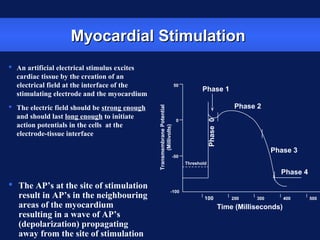
Basic Technical Concepts In Cardiac Pacing Ppt

30 Thousand Circulatory Royalty Free Images Stock Photos Pictures Shutterstock
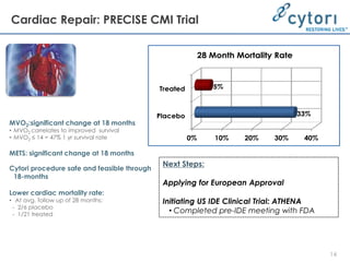
Stem Cell Meeting On The Mesa 2011 Cytx Presentation Ppt
![]()
Cardiac Muscle Tissue Function And Labeled Diagram Getbodysmart
Muscle The Histology Guide
Which Organelle Would Be Abundant In Skeletal Muscle And Palisade Muscle Quora
Why Does Cardiac Muscle Tissue Have Large Numbers Of Mitochondria Quora
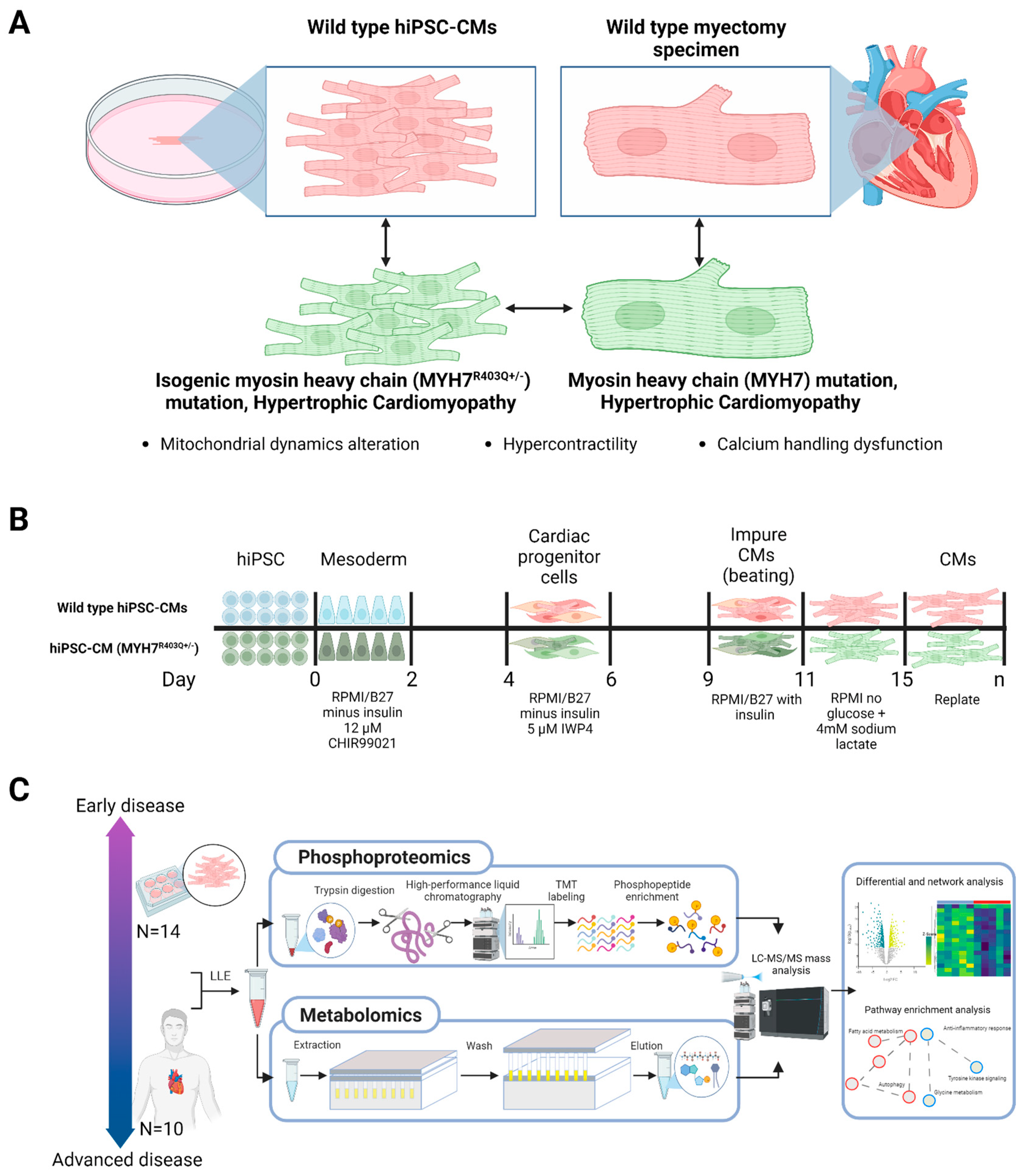
Ijms Free Full Text Multi Omics Profiling Of Hypertrophic Cardiomyopathy Reveals Altered Mechanisms In Mitochondrial Dynamics And Excitation Ndash Contraction Coupling
Cardiac Muscle Cell Hi Res Stock Photography And Images Alamy
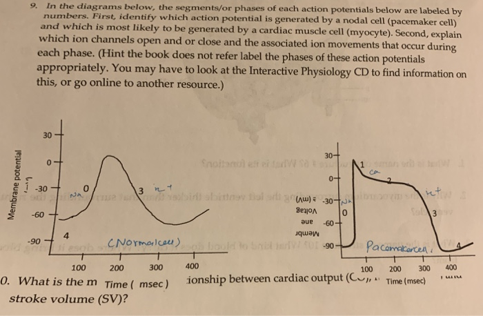
Solved 9 In The Diagrams Below The Segments Or Phases Of Chegg Com
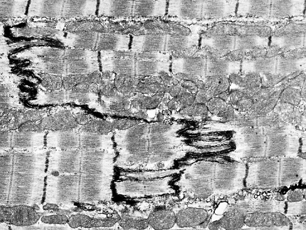
Cardiac Muscle Cell Em
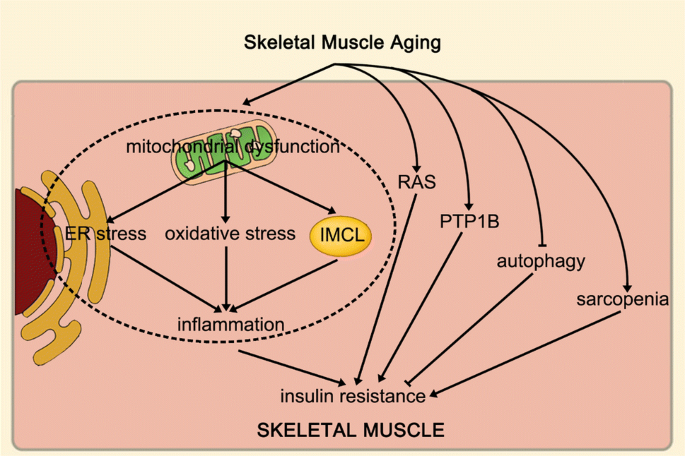
Mechanism Of Increased Risk Of Insulin Resistance In Aging Skeletal Muscle Diabetology Metabolic Syndrome Full Text

Cardiac Muscle Definition Function And Structure Biology Dictionary